
From discovery to validation: Six applications of peptide microarrays in immuno-oncology
Article
Immuno-oncology is undergoing a revolution, driven by therapies that harness and enhance the power of the immune system to fight cancer. As immune checkpoint inhibitors, therapeutic antibodies, and personalized vaccines become more widespread, the urgent need for precise characterization of immune responses and accurate identification of therapeutic targets has never been clearer. At the heart of this precision lies peptide microarrays, a high-throughput, versatile technology capable of revealing detailed interactions between antibodies, antigens, and immune receptors at the level of single amino acid resolution.
In recent years, peptide microarrays have moved from being niche research tools to essential platforms in immuno-oncology research, diagnostics, and therapeutic development. Their ability to rapidly map epitopes, profile antibody responses, and discover predictive biomarkers has opened unprecedented opportunities to refine cancer immunotherapies and maximize patient outcomes.
In this article, we highlight six innovative applications of peptide microarrays that are pushing the boundaries of immuno-oncology, from pinpointing novel tumor antigens and predicting patient responses, to guiding the design of next-generation therapeutics.
1. Discovering a new antibody-drug conjugate targeting prostate cancer
Overview: Scientists at the University of Pittsburgh Medical School have recently developed a novel therapeutic approach by engineering a fully human monoclonal antibody (named F12) targeting prostate stem cell antigen (PSCA), a cell-surface protein abundantly expressed in prostate tumors. Using phage display and subsequent structure-based affinity maturation, the researchers developed an antibody with high affinity and specificity, demonstrated through extensive binding studies. They further developed this antibody into an antibody-drug conjugate (ADC) by linking it to the potent antimitotic agent monomethyl auristatin E (MMAE). The resulting ADC showed robust anti-tumor activity in vitro and demonstrated dose-dependent tumor growth inhibition in a human prostate cancer xenograft mouse model.
Why this matters: Prostate cancer is a leading cause of cancer-related death, particularly due to its progression to androgen-independent and metastatic forms, which lack effective treatments. PSCA, widely expressed in prostate cancer tissues with minimal expression in normal tissues, presents an ideal therapeutic target. Previous antibody-based therapies targeting PSCA faced limited clinical success due to issues such as antigen heterogeneity and tumor microenvironment-related challenges. This research offers a highly specific and fully human antibody-ADC approach, which not only targets cancer cells directly but also addresses limitations seen in other therapeutic antibodies. If successfully translated into clinical practice, it could substantially enhance treatment outcomes for prostate cancer patients and potentially extend to other PSCA-expressing solid tumors.
How peptide microarrays were used: PEPperPRINT's peptide microarray was used to precisely map the binding epitope of the newly identified antibody F12 on the PSCA protein. The method involved generating cyclic peptides representing different overlapping sequences of the PSCA antigen. By screening these peptides with antibody F12, researchers identified a specific epitope that the antibody binds with high specificity. This detailed mapping confirmed the precision and selectivity of antibody F12, providing critical validation that enhanced confidence in its therapeutic potential and guided its further development as an ADC.
Reference: Chu, X. et al. “Discovery of a novel highly specific, fully human PSCA antibody and its application as an antibody-drug conjugate in prostate cancer.” mAbs. (2024 August). https://doi.org/10.1080/19420862.2024.2387240
2. Identifying a new bispecific antibody for the treatment of CD30-positive cancers
Overview: In this study, a team of researchers from various North American universities developed novel anti-CD30/CD3 bispecific antibodies (biAbs) to directly engage human T cells and redirect their potent cytotoxic effects toward CD30-positive tumor cells. The study identified two promising antibodies, distinct from current clinical antibodies, through careful screening for binding specificity, affinity, and unique epitope targeting. The biAbs, when loaded onto activated human T cells, demonstrated strong tumor cell binding, robust cytokine production, and effective killing of CD30-expressing tumor cells in vitro and significantly prolonged survival in a mouse xenograft model.
Why this matters: CD30 is widely recognized as an attractive target for immunotherapy, as it is expressed in several cancers, including Hodgkin lymphoma and certain non-Hodgkin lymphomas, but shows limited expression in normal tissues. Previous ADCs, such as brentuximab vedotin, have shown promise but have limitations, like toxicity and therapy resistance. This study's approach of using bispecific antibodies also leverages the natural killing capacity of T cells while avoiding the genetic modification complexities associated with CAR-T cell therapies. It represents an important advancement toward safer and potentially more effective treatments for refractory and relapsed CD30-positive cancers. The demonstrated specificity, potency, and ability of these bispecific antibodies to redirect T cells against tumor cells could significantly impact therapeutic outcomes and patient survival, offering new hope to patients with few therapeutic options.
How peptide microarrays were used: PEPperPRINT’s peptide microarrays played a key role in precisely mapping the linear epitopes recognized by the newly developed anti-CD30 antibodies. The microarrays included hundreds of overlapping linear peptides spanning the CD30 protein. Researchers screened the lead antibody against these arrays to identify specific peptide sequences it recognized, revealing a unique epitope distinct from that of brentuximab vedotin. This detailed epitope mapping provided critical validation of antibody specificity and helped differentiate their antibodies from existing clinical candidates. Such precise characterization ensures confidence in the antibody's unique mechanism of action, potentially reducing cross-reactivity risks and improving therapeutic outcomes.
Reference: Faber, Mary L. et al. “Novel anti-CD30/CD3 bispecific antibodies activate human T cells and mediate potent anti-tumor activity.” Frontiers in Immunology. (2023 August). https://doi.org/10.3389/fimmu.2023.1225610
3. Measuring B cell antibody responses in breast cancer immunotherapy
Overview: In this study, researchers at the University of North Carolina School of Medicine investigated how antibody-producing B cells respond to tumor antigens in patients with metastatic triple-negative breast cancer (TNBC) undergoing immunotherapy. Patients received cyclophosphamide, a chemotherapy agent that reduces regulatory immune cells, followed by pembrolizumab, an immune checkpoint inhibitor designed to enhance immune attack on cancer cells. The team used computational methods and custom peptide microarrays to evaluate antibody responses to both mutated tumor-specific neoantigens and normal self-antigens. Although only a subset of predicted neoantigens elicited measurable antibody responses, strikingly varied patient-specific patterns emerged. Importantly, a significant increase—or "boostability"—in antibody response was observed following treatment, especially prominent in one patient who achieved a complete clinical response. This patient's antibodies strongly targeted a self-epitope within the EPS8 protein, a known oncogenic factor in breast cancer, highlighting a potentially crucial role for self-reactive B cells in successful immunotherapy outcomes.
Why this matters: TNBC is one of the most aggressive and difficult-to-treat breast cancers, lacking effective targeted therapies and often resisting standard immunotherapies. Understanding the role of B cells and tumor-specific antibodies in cancer immunotherapy is crucial, as it may reveal novel mechanisms of tumor immunity and potential biomarkers of therapeutic efficacy. Previous research primarily emphasized the role of T cells in immune checkpoint therapies; however, emerging evidence shows that B cell responses significantly impact clinical outcomes. Identifying antibody targets associated with successful immunotherapy responses could inform future personalized therapeutic strategies, vaccine development, and biomarker discovery in cancers like TNBC, which currently has limited effective treatments.
How peptide microarrays were used: PEPperPRINT’s peptide microarrays were central to this study's methodology. Researchers generated custom peptide microarrays featuring thousands of predicted linear peptides, including both mutated neoantigens and their corresponding unaltered self-peptides. Patient plasma samples collected before and after pembrolizumab treatment were incubated on these microarrays. Bound antibodies were visualized using fluorescently labeled secondary antibodies. The technique allowed precise identification of peptides eliciting specific antibody responses and measurement of the intensity and specificity of these responses.
Reference: Routh, ED. et al, “Evaluation of tumor antigen-specific antibody responses in patients with metastatic triple negative breast cancer treated with cyclophosphamide and pembrolizumab”, Journal for ImmunoTherapy of Cancer. (2023 March). https://doi.org/10.1136/jitc-2022-005848
4. Using IgM antibody profiles as novel biomarkers in brain tumor diagnosis
Overview: This research, led by scientists from Bulgaria, explored whether patterns of immunoglobulin M (IgM) antibody responses in patients with brain tumors, particularly glioblastoma multiforme (GBM) and brain metastases, differ from those in non-tumor patients. Researchers utilized a peptide microarray featuring thousands of overlapping linear peptides derived from known tumor-associated antigens (TAAs) and cancer-related viral epitopes. By mapping IgM antibody reactivities from patient sera onto a specially developed "reactivity graph," the team was able to analyze complex patterns of antibody cross-reactivity. This approach revealed specific clusters of peptides whose IgM binding patterns were significantly associated with particular tumor diagnoses.
Why this matters: The immune system continuously produces IgM, antibodies typically known for their polyspecificity and natural autoreactivity. Current cancer diagnostics often focus on IgG antibodies or direct tumor antigen detection, neglecting the diagnostic potential of the natural IgM antibody repertoire. IgM antibodies reflect fundamental immune processes, such as self-recognition, blood group antigen reactions, and responses to viral antigens. By identifying distinct IgM antibody signatures linked to specific tumor types, the research demonstrates how IgM profiling could serve as a novel, non-invasive biomarker approach for cancer diagnosis and patient stratification. If validated further, this method could enable earlier detection and improved monitoring of solid tumors, potentially revolutionizing the clinical use of immunological biomarkers.
How peptide microarrays were used: PEPperPRINT’s peptide microarrays provided a platform to screen IgM antibodies against thousands of defined linear epitopes derived from both human tumor antigens and cancer-related viruses. The array included peptides designed as overlapping sequences to ensure precise epitope mapping. Patient sera from GBM, brain metastases, and non-tumor controls were incubated on these microarrays, and IgM binding patterns were analyzed and quantified. Through this approach, the researchers successfully differentiated diagnostic groups based on specific IgM reactivity patterns, establishing a proof-of-concept for using IgM immunoprofiling as a diagnostic tool.
Reference: Ferdinandov, Dilyan, et al. “Reactivity Graph Yields Interpretable IgM Repertoire Signatures as Potential Tumor Biomarkers.” International Journal of Molecular Sciences. (2023 January). https://doi.org/10.3390/ijms24032597
5. Identifying a marker of drug resistance to anti-VEGF therapy in melanoma
Overview: To investigate potential mechanisms underlying resistance to anti-vascular endothelial growth factor (VEGF) therapy, a team of researchers in Argentina explored the role of galectin-1 (Gal1), a glycan-binding protein with known proangiogenic and immunomodulatory properties. Using clinical data from melanoma patients enrolled in the AVAST-M trial, researchers found that circulating plasma Gal1 levels increased specifically in patients treated with bevacizumab who experienced recurrence and poorer survival outcomes. They demonstrated that Gal1 actively promotes angiogenesis even when VEGF is blocked. A newly developed fully human anti-Gal1 monoclonal antibody effectively neutralized circulating Gal1 activity, reducing endothelial cell migration, tubulogenesis, and VEGFR2 activation. This confirmed that Gal1 contributes to adaptive resistance to anti-VEGF treatments.
Why this matters: While anti-VEGF therapies such as bevacizumab initially suppress tumor growth by inhibiting angiogenesis, many tumors eventually develop adaptive resistance mechanisms, resuming growth despite treatment. Identifying and targeting these resistance pathways is critical to improving clinical outcomes. The study provides strong clinical evidence that Gal1 functions as a compensatory mechanism allowing tumors to sustain angiogenesis and survive VEGF blockade. This highlights the potential of Gal1 as both a predictive biomarker for resistance to anti-VEGF therapies and as a novel therapeutic target.
How peptide microarrays were used: PEPperPRINT’s peptide microarrays were crucial for identifying and characterizing the binding epitopes of the newly developed fully human anti-Gal1 antibodies. The microarray analysis involved screening antibodies against hundreds of overlapping peptides derived from the Gal1 protein, enabling the precise determination of the antibodies' specific binding sites. This high-resolution epitope mapping revealed a unique epitope recognized by the antibodies. Confirming this specificity was essential for establishing the antibodies' effectiveness and selectivity, ensuring they could specifically neutralize Gal1 without cross-reactivity to other related galectins.
Reference: Bannoud, Nadia, et al. “Circulating galectin-1 delineates response to bevacizumab in melanoma patients and reprograms endothelial cell biology.” Proceedings of the National Academy of Sciences. (2023 January). https://doi.org/10.1073/pnas.2214350120
6. Monitoring PD-L1 antibody effectiveness with non-invasive imaging
Overview: Accurate identification of PD-L1 expression in tumors is critical for selecting appropriate patients for therapy. A team of scientists at the Mayo Clinic in the US addressed this challenge by developing a novel imaging approach: a non-invasive immunoPET technique using zirconium-89 labeled antibodies targeting PD-L1. Researchers compared a newly developed anti-PD-L1 antibody (anti-PD-L1-B11), which binds PD-L1 at a distinct epitope, against the therapeutic antibody atezolizumab. Utilizing preclinical breast cancer and melanoma models, anti-PD-L1-B11 radiolabeled with zirconium-89 demonstrated superior imaging properties. This antibody exhibited high specificity and affinity for PD-L1-positive tumor cells and showed favorable biodistribution and tumor uptake characteristics, even in the presence of therapeutic doses of atezolizumab.
Why this matters: While immune checkpoint inhibitors targeting PD-1/PD-L1 pathways have revolutionized cancer therapy, only a subset of patients responds effectively. Current clinical methods for assessing tumor PD-L1 expression—primarily immunohistochemistry—are invasive, susceptible to sampling errors, and unable to capture tumor heterogeneity or changes in PD-L1 expression over time. Non-invasive immunoPET imaging overcomes these limitations by enabling real-time assessment of PD-L1 status across entire tumors and metastases. This approach could significantly improve patient selection, treatment planning, and therapy monitoring for PD-1/PD-L1 targeted immunotherapies. By employing an antibody targeting a different binding site than therapeutic antibodies, this novel method also avoids competition from therapeutic doses, making it highly practical for clinical settings and potentially enhancing treatment efficacy and patient outcomes.
How peptide microarrays were used: PEPperPRINT’s peptide microarray technology was used to precisely map and differentiate the binding epitopes of the anti-PD-L1-B11 antibody from those of atezolizumab. This epitope mapping involved screening the antibodies against overlapping peptides covering the entire PD-L1 protein sequence. The analysis confirmed that anti-PD-L1-B11 binds to a distinct PD-L1 epitope compared to atezolizumab, thus validating the antibody's unique specificity and suitability for use alongside therapeutic antibodies without competitive interference. This crucial step reinforced the potential clinical utility of anti-PD-L1-B11 as a superior PET imaging probe.
Reference: Bansal, Aditya, et al. “Non-invasive immunoPET imaging of PD-L1 using anti-PD-L1-B11 in breast cancer and melanoma tumor model.” Nuclear Medicine and Biology. (2021 May). https://doi.org/10.1016/j.nucmedbio.2021.05.004
How peptide microarrays can support your research
With unmatched precision, flexibility, and throughput, PEPperPRINT helps researchers rapidly map antibody epitopes, profile immune responses, and identify robust biomarkers crucial for advancing personalized cancer treatments.
Whether your research involves epitope characterization for novel therapeutic antibodies, screening patient sera for predictive biomarkers, or mapping immune responses to emerging tumor antigens, PEPperPRINT’s platform delivers clarity, confidence, and actionable data.
Contact PEPperPRINT today to discuss how peptide microarrays can unlock deeper insights and drive your immuno-oncology research to the newt level.
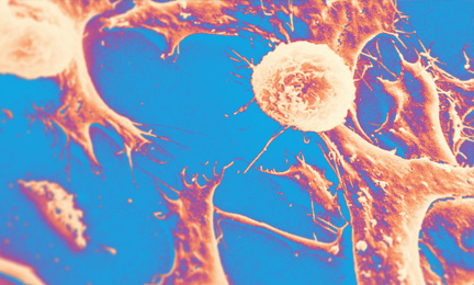
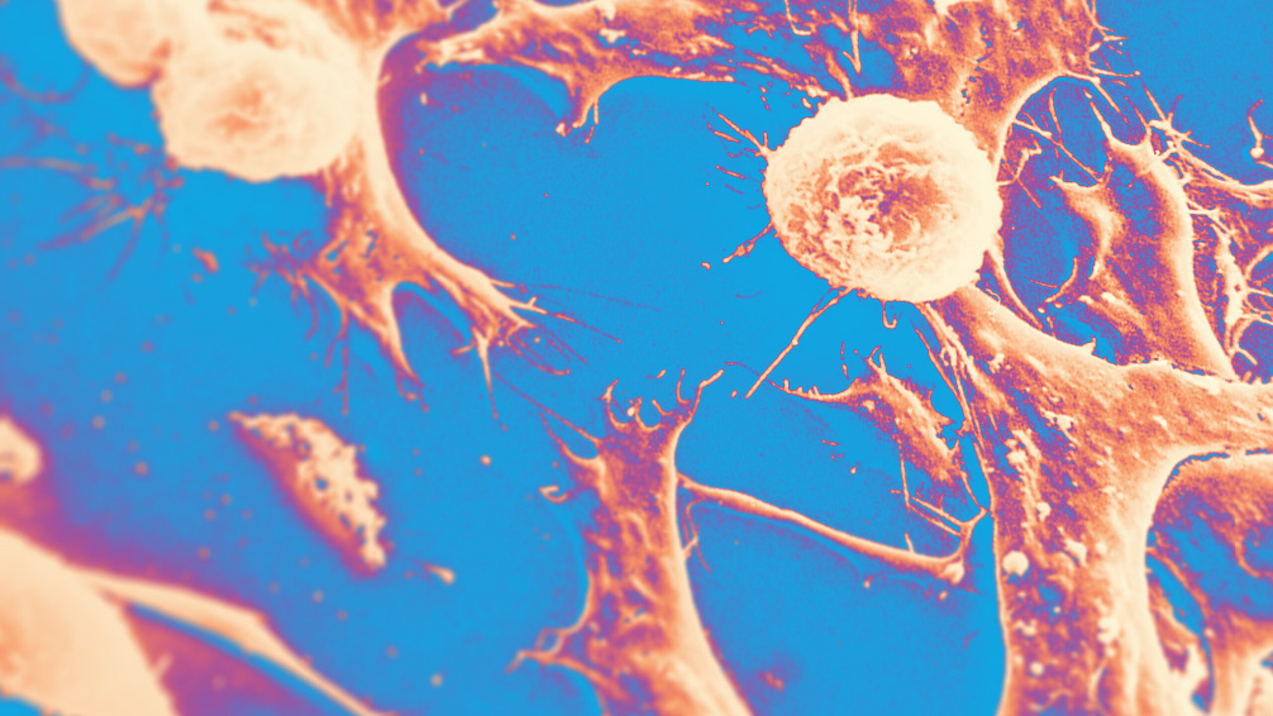
recolored electron micrograph of Wilms' tumor (original image from the National Cancer Institute)